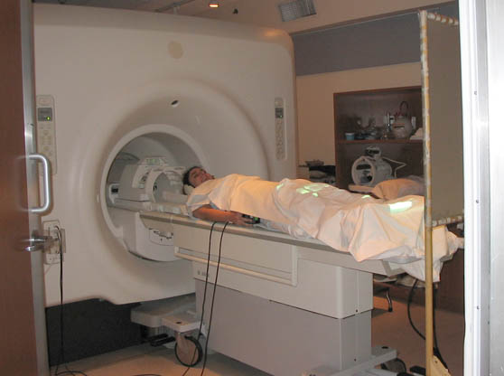
Yes that's me, going in for fMRI when I volunteered for a neurology study at Sunnybrook
hospital. I was in there for several hours on several occasions. I made a lot of notes
on what it's like to be inside the scanner, but we'll save that for another day.
I'm auditing a class called "Introduction to fMRI." Disclaimer in case anyone winds up here because they are looking for actual neuroscience: I'm not a scientist and I don't have a fucking clue what I am doing!
Our teacher says that the MRI scanner is not a black box. He wants his students to be able to explain how it works. So here goes, in my own words. If you have any self respect, you will not quote me on this.
MRI stands for Magnetic Resonance Imaging. It used to be called NMR, Nuclear Magnetic Resonance, but they changed the name in the 70s because people at the time were so freaked out by nuclear technology of any kind. An MRI scan produces a static, anatomical image of the brain. The 'f' in fMRI stands for functional. fMRI scans are series of static images that show changes in brain activity over a period of time (like animated gifs).
Anatomical MRI scans are usually at a higher resolution than fMRI scans, because the scan itself can take longer. The basic unit in these digital images is a voxel, which is like a 3D version of a pixel. Voxels can be bigger (low rez) or smaller (high rez) depending on how much time you take.
Okay. 1) How do you use magnets and radio frequencies to collect data about someone's brain? 2) How do you specify exactly what you want to measure within the brain? 3) How do you translate your data into an image, or series of images? I can almost barely manage to discuss the answer to 1; I have even less of a grip on 2; we're just getting started on 3 and so far it is just plain mind-boggling. Shall we begin?
1) The scanner contains a really big, really powerful magnetic coil that is always on. When someone's head is inside this static magnetic field, the protons in their hydrogen atoms tend to "spin" together in one direction. Spin is a quantum term and doesn't really mean spin like we experience it. This is the bit where Richard Feynman would say something like "if you think you understand it you don't understand it." So just think of spin as a bit of a metaphor, but one that's fairly apt. Anyhow, a lot of the protons get lined up (the stronger the field, the more alignment). This is the state that they need to be in to get measured.
The scanner also contains radio frequency (RF) coils that can be turned on and off quickly. Once the protons are lined up because of the magnet, the RF kicks in and flips some of them into higher energy states. When the RF is switched off, the protons flip back down to the lower energy state. This switching back down is a release of energy (the exact same amount of energy that the RF coils introduced). This means that each proton that is shifting releases a photon, create a radio frequency. The scanner also has a set of RF detector coils that pick up the frequency and thereby notice when the photons are released. That's the data. (Some scanners use the same RF coils to both induce and receive the frequencies, some have separate coils, but in either case the RF is always the same coming out as going in).
2) This bit is super technical and I'm unable to fully digest it. Here's what I've got. The magnetic and RF fields are not uniform, but gradient, meaning that they change along a spatial plane. So different frequencies can be correlated with different physical locations, ie: the field is passing through somebody's physical head, and the field is different, and producing different effects, in different areas of the brain. As mentioned above, everything happens within the very strong, constant static magnetic field.
In order to isolate a slice of the brain you need to set the RF field that you turn on and off to a specific range of frequencies. That way the only information coming back at you will have to be from the slice that resonates at that range of frequencies. That's 1 dimension. In order to get 2 more dimensions you need to do 2 more things. One is that you introduce another field in order to make the changes in the protons' spin dependent on their location. This is called frequency encoding. I don't get how or why this works, but what frequency encoding does is give you another dimension.
So now you've got both a horizontal slice, and a vertical range of behaviours across that slice. The other thing you do is called phase encoding. This involves applying yet another field, perpendicular to the other two. I'm not sure, but I think this bit is temporal. So for phase encoding you apply a bunch of different fields in sequence and watch how the protons change. Anyhow. Once you do slice selection, frequency encoding, and phase encoding you get specific 3D data.
One important piece of this is a mathematical thingy called the Fourier Transform. Again, I'm in way over my head, but as I understand it, the data that's coming out of the scanner is like a resonant harmonic of all the various frequencies combined into one sine wave. Fourier Transform is a way of looking at that wave and breaking it down into its component frequencies. Or vice versa, or both. I think you can use Fourier Transform to combine frequencies into a single wave and to break them out of a single wave.
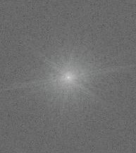
3) Okay. Now that I'm totally lost, on completely shaky ground, let me continue. The first representation of the data, before it shows up in "image space" is in "k-space." Whoo! K-space is cool and it looks like some kind of cosmic event. K-space contains all the spatial frequency data from the scan.
Depending on what sections of k-space you draw information from, you will get different images of the brain at different contrasts, at different resolutions, and/or in different segments. In our next class we will be learning about "the sequence of events (gradients, RF) used to traverse k-space in order to read an entire image." If I can find my way back out, I'll try to make a report.
|
This is SO interesting! You're doing us all a big favour, laying it out like this.
I agree, and if I had a brain tumor I'd prefer to get the bad news from a good friend, not some fucking "doctor". GO SALLY GO!
LOL.
Here's a reputable document:
Weishaupt, Dominick, et.al., How Does MRI Work? An Introduction to the Physics and Function of Magenetic Resonance Imaging, (Berlin & New York : Springer, 2006)
Coo-ehl. Rob just sent me a link to DIY EEG!
The neurological study I volunteered for had an EEG component that was, in some ways, even weirder than the MRI. (When they put the electrodes all over my skull with wires coming out I momentarily freaked because I had forgotten that the electricity being measured was already in my brain.)
As long as they mounted a Scratch Sally first, it's all good.
I have to say that based on the tiny bit of information provided in that link I'm a bit skeptical about the DIY EEG. The participants are moving their face muscles to induce different brain waves. But when I was on the EEG they told me to blink as little as possible because the electrodes would pick up signals from that activity that interferred with the reading of the actual brain waves. I'm afraid that the Arduino EEG Project is only picking up signals from the face, and not the brain itself. But maybe they've accounted for that - it looks like a competely different kind of interface than the one I experienced.
I googled scratch sally. I found this: "I used a Bamboo pole as an arm extension to lightly scratch Sally around the withers and down her back, without having to get too close to her. [...] Sally never once tried to bite or kick me, even when she was really scared." (we're good that way.)
It all makes perfect sense to me, but what the hell is an animated gif?
meanwhile...
"Two months later, classmates of the Shipleys' daughter, Shannon, who was also injured in the accident, took a field trip to the Richmond County Mortuary. Several students noticed a jar holding a human brain suspended in formaldehyde.
"In what can only be described as a surreal coincidence," Mastro wrote, "the label on the jar indicated that the brain was that of Jesse Shipley, a circumstance which evoked strong emotional reactions from some of the students who were present."
http://www.law.com/jsp/law/LawArticleFriendly.jsp?id=1202472788415
omg, that's an awful story.
I was looking closely at an anatomical diagram of a brain the other day and for the first time (yes I am a bit slow when it comes to really obvious physiological reactions) I felt the distinct and creepy feeling that it is really just not designed to be detached from the body. Like when you cut yourself badly and see "inside" stuff that just isn't supposed to be seen. It's sort of funny considering how very very often the brain is represented graphically. How many times have you seen a t-shirt with an image of a liver on it?
This contains many incorrect statements - if you are interested...
The word nuclear was not dropped because people freaked out - it was a radiology turf war and they wanted to keep NMR apart from nuclear medicine.
Protons do not get "lined up" protons exist in one of two energy states and their energy state is all that changes. What gets affected is their net magnetic vector - it is this vector that is manipulated, not the physical orientation of the nuclei.
Although RF is used to resonate the hydrogen, the receiver coil measures the resulting rotating net magnetic vector via electromagnetic induction. Not photons. The "RF coming out" is therefore negligible in comparison to the RF "going in"
The primary magnetic field and the RF are VERY uniform.
You are thinking about the spatially varying gradient fields that are applied separately to encode the signal.
Your other interpretations are very good!
|
Yes that's me, going in for fMRI when I volunteered for a neurology study at Sunnybrook
hospital. I was in there for several hours on several occasions. I made a lot of notes
on what it's like to be inside the scanner, but we'll save that for another day.
I'm auditing a class called "Introduction to fMRI." Disclaimer in case anyone winds up here because they are looking for actual neuroscience: I'm not a scientist and I don't have a fucking clue what I am doing!
Our teacher says that the MRI scanner is not a black box. He wants his students to be able to explain how it works. So here goes, in my own words. If you have any self respect, you will not quote me on this.
MRI stands for Magnetic Resonance Imaging. It used to be called NMR, Nuclear Magnetic Resonance, but they changed the name in the 70s because people at the time were so freaked out by nuclear technology of any kind. An MRI scan produces a static, anatomical image of the brain. The 'f' in fMRI stands for functional. fMRI scans are series of static images that show changes in brain activity over a period of time (like animated gifs).
Anatomical MRI scans are usually at a higher resolution than fMRI scans, because the scan itself can take longer. The basic unit in these digital images is a voxel, which is like a 3D version of a pixel. Voxels can be bigger (low rez) or smaller (high rez) depending on how much time you take.
Okay. 1) How do you use magnets and radio frequencies to collect data about someone's brain? 2) How do you specify exactly what you want to measure within the brain? 3) How do you translate your data into an image, or series of images? I can almost barely manage to discuss the answer to 1; I have even less of a grip on 2; we're just getting started on 3 and so far it is just plain mind-boggling. Shall we begin?
1) The scanner contains a really big, really powerful magnetic coil that is always on. When someone's head is inside this static magnetic field, the protons in their hydrogen atoms tend to "spin" together in one direction. Spin is a quantum term and doesn't really mean spin like we experience it. This is the bit where Richard Feynman would say something like "if you think you understand it you don't understand it." So just think of spin as a bit of a metaphor, but one that's fairly apt. Anyhow, a lot of the protons get lined up (the stronger the field, the more alignment). This is the state that they need to be in to get measured.
The scanner also contains radio frequency (RF) coils that can be turned on and off quickly. Once the protons are lined up because of the magnet, the RF kicks in and flips some of them into higher energy states. When the RF is switched off, the protons flip back down to the lower energy state. This switching back down is a release of energy (the exact same amount of energy that the RF coils introduced). This means that each proton that is shifting releases a photon, create a radio frequency. The scanner also has a set of RF detector coils that pick up the frequency and thereby notice when the photons are released. That's the data. (Some scanners use the same RF coils to both induce and receive the frequencies, some have separate coils, but in either case the RF is always the same coming out as going in).
2) This bit is super technical and I'm unable to fully digest it. Here's what I've got. The magnetic and RF fields are not uniform, but gradient, meaning that they change along a spatial plane. So different frequencies can be correlated with different physical locations, ie: the field is passing through somebody's physical head, and the field is different, and producing different effects, in different areas of the brain. As mentioned above, everything happens within the very strong, constant static magnetic field.
In order to isolate a slice of the brain you need to set the RF field that you turn on and off to a specific range of frequencies. That way the only information coming back at you will have to be from the slice that resonates at that range of frequencies. That's 1 dimension. In order to get 2 more dimensions you need to do 2 more things. One is that you introduce another field in order to make the changes in the protons' spin dependent on their location. This is called frequency encoding. I don't get how or why this works, but what frequency encoding does is give you another dimension.
So now you've got both a horizontal slice, and a vertical range of behaviours across that slice. The other thing you do is called phase encoding. This involves applying yet another field, perpendicular to the other two. I'm not sure, but I think this bit is temporal. So for phase encoding you apply a bunch of different fields in sequence and watch how the protons change. Anyhow. Once you do slice selection, frequency encoding, and phase encoding you get specific 3D data.
One important piece of this is a mathematical thingy called the Fourier Transform. Again, I'm in way over my head, but as I understand it, the data that's coming out of the scanner is like a resonant harmonic of all the various frequencies combined into one sine wave. Fourier Transform is a way of looking at that wave and breaking it down into its component frequencies. Or vice versa, or both. I think you can use Fourier Transform to combine frequencies into a single wave and to break them out of a single wave.
3) Okay. Now that I'm totally lost, on completely shaky ground, let me continue. The first representation of the data, before it shows up in "image space" is in "k-space." Whoo! K-space is cool and it looks like some kind of cosmic event. K-space contains all the spatial frequency data from the scan.
Depending on what sections of k-space you draw information from, you will get different images of the brain at different contrasts, at different resolutions, and/or in different segments. In our next class we will be learning about "the sequence of events (gradients, RF) used to traverse k-space in order to read an entire image." If I can find my way back out, I'll try to make a report.
- sally mckay 9-30-2010 1:12 pm
This is SO interesting! You're doing us all a big favour, laying it out like this.
- M.Jean 9-30-2010 2:26 pm
I agree, and if I had a brain tumor I'd prefer to get the bad news from a good friend, not some fucking "doctor". GO SALLY GO!
- L.M. 9-30-2010 2:32 pm
LOL.
- sally mckay 9-30-2010 3:37 pm
Here's a reputable document:
Weishaupt, Dominick, et.al., How Does MRI Work? An Introduction to the Physics and Function of Magenetic Resonance Imaging, (Berlin & New York : Springer, 2006)
- sally mckay 9-30-2010 3:43 pm
Coo-ehl. Rob just sent me a link to DIY EEG!
The neurological study I volunteered for had an EEG component that was, in some ways, even weirder than the MRI. (When they put the electrodes all over my skull with wires coming out I momentarily freaked because I had forgotten that the electricity being measured was already in my brain.)
- sally mckay 10-01-2010 4:29 am
As long as they mounted a Scratch Sally first, it's all good.
- rob (guest) 10-01-2010 4:38 pm
I have to say that based on the tiny bit of information provided in that link I'm a bit skeptical about the DIY EEG. The participants are moving their face muscles to induce different brain waves. But when I was on the EEG they told me to blink as little as possible because the electrodes would pick up signals from that activity that interferred with the reading of the actual brain waves. I'm afraid that the Arduino EEG Project is only picking up signals from the face, and not the brain itself. But maybe they've accounted for that - it looks like a competely different kind of interface than the one I experienced.
- sally mckay 10-01-2010 4:48 pm
I googled scratch sally. I found this: "I used a Bamboo pole as an arm extension to lightly scratch Sally around the withers and down her back, without having to get too close to her. [...] Sally never once tried to bite or kick me, even when she was really scared." (we're good that way.)
- sally mckay 10-01-2010 5:03 pm
It all makes perfect sense to me, but what the hell is an animated gif?
- joester (guest) 10-01-2010 8:03 pm
meanwhile...
"Two months later, classmates of the Shipleys' daughter, Shannon, who was also injured in the accident, took a field trip to the Richmond County Mortuary. Several students noticed a jar holding a human brain suspended in formaldehyde.
"In what can only be described as a surreal coincidence," Mastro wrote, "the label on the jar indicated that the brain was that of Jesse Shipley, a circumstance which evoked strong emotional reactions from some of the students who were present."
http://www.law.com/jsp/law/LawArticleFriendly.jsp?id=1202472788415
- joester (guest) 10-02-2010 4:09 pm
omg, that's an awful story.
I was looking closely at an anatomical diagram of a brain the other day and for the first time (yes I am a bit slow when it comes to really obvious physiological reactions) I felt the distinct and creepy feeling that it is really just not designed to be detached from the body. Like when you cut yourself badly and see "inside" stuff that just isn't supposed to be seen. It's sort of funny considering how very very often the brain is represented graphically. How many times have you seen a t-shirt with an image of a liver on it?
- sally mckay 10-02-2010 4:54 pm
- sally mckay 10-02-2010 5:15 pm
- sally mckay 10-02-2010 5:30 pm
This contains many incorrect statements - if you are interested...
The word nuclear was not dropped because people freaked out - it was a radiology turf war and they wanted to keep NMR apart from nuclear medicine.
Protons do not get "lined up" protons exist in one of two energy states and their energy state is all that changes. What gets affected is their net magnetic vector - it is this vector that is manipulated, not the physical orientation of the nuclei.
Although RF is used to resonate the hydrogen, the receiver coil measures the resulting rotating net magnetic vector via electromagnetic induction. Not photons. The "RF coming out" is therefore negligible in comparison to the RF "going in"
The primary magnetic field and the RF are VERY uniform.
You are thinking about the spatially varying gradient fields that are applied separately to encode the signal.
Your other interpretations are very good!
- anonymous (guest) 3-06-2020 7:17 am