|
Lorna Mills and Sally McKay
Digital Media Tree this blog's archive OVVLvverk Lorna Mills: Artworks / Persona Volare / contact Sally McKay: GIFS / cv and contact |
View current page
...more recent posts
The Talairach Atlas
| Wikipedia says: "By defining standard anatomical landmarks that could be identified on different subjects (the anterior and posterior commissures), it became easier to spatially warp an individual brain image obtained through Magnetic Resonance Imaging (MRI), Positron Emission Tomography (PET) and other imaging methods to this 'standard Talairach space'. One can then make inferences about tissue identity at a specific location by referring to the atlas." Everything2 says:" There are some problems with the Talairach atlas. First of all, the atlas is based on one brain, which happens to be the brain of a 60-year-old French woman. Only one hemisphere was mapped, assuming that the hemispheres were symmetrical (although as a rule they are not). The most notable difference between the Talairach brain and other brains is its size. The Talairach brain is considerably smaller than the average brain by up to 10 millimeters in each dimension. Because brain sections are typically taken every 2 millimeters, this leaves a total of 15 slices that would go unnaccounted for. Also, the Talairach atlas leaves out the majority of the brain stem and cerebellum." |
9 different people's brains, aligned at the thalamus.
ff
A few weeks ago I made a post about fMRI. This is another one. I'm currently auditing a class called "Introduction to fMRI." Disclaimer in case anyone winds up here because they are looking for actual neuroscience: I'm not a scientist and I don't have a fucking clue what I am doing! I'm trying my best, but I suggest you do not quote me.  Detail from autopsy photo of a human brain at Wikimedia (cropped and enlarged). fMRI is a digital imaging challenge extraordinaire. Two of the biggest concerns are resolution and signal to noise ratio. The image above is a photograph showing some of the vasculature on the surface of the brain. fMRI measures something called the blood-oxygen-level dependent (BOLD) effect. In very simple terms, when synaptic action happens between neurons the process requires energy, and blood provides the fuel. So when a certain area of the brain is busy, blood will concentrate there. The nuclear processes ("spinning" protons) activated and detected by the scanner happen in the blood flow that correlates to the synaptic activity, not in the neurons themselves. Happily, the network of veins and arteries is extremely fine and precise — maintaining a discrete structure right down to level of individual blood vessels — so the vasculature itself doesn't limit the resolution of the scan. The BOLD effect takes place over time, as blood flows in and out. The signal tends to look like this. 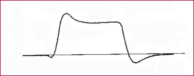 And this is why. 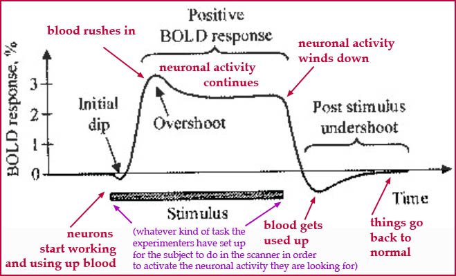 The useful amount of signal is limited by the time scale of the BOLD response. Also, there are issues of pooling in nearby veins when the blood recedes that can cause noise. Deciding on resolution is a balancing act. The smaller you make your voxels the more precisely you can localise the effects, but you will also have a weaker signal because is less blood to measure per unit. And you want a strong signal because there can be a TON of noise. Head movements cause a lot of noise, as well as inconsistencies in the magnetic field that can be caused by the scanner itself, the morphology of the person's brain, and thingies in the blood. It's very easy to get false positives. So the size of your voxels needs to be large enough that you get a strong signal, but small enough that the signal isn't completely full of junk. The scanner detects the entire electro-magnetic signal coming from each voxel. You might be looking for something like this. 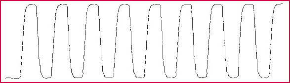
But you are quite likely to get something more like this. 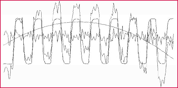 Which means its pretty easy to wind up with an fMRI image showing false positives unless you are very careful with your data. 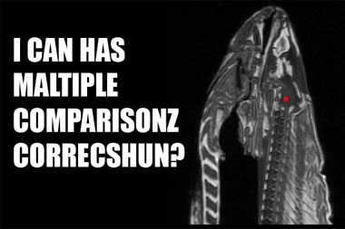
For more on Craig Bennett's dead salmon, see this post from last year. Also, of course, different brain activities take different amounts of time. So you also need to consider how long your activation signal will be, based on the experiment. Some brain processes that take more than a few seconds to happen just aren't that amenable to being measured by fMRI. And then there are the problems of "normalization" across different test subjects. Everybody's brain is different. So some noise is going to enter into your data when you start trying to combine information from different people into one stream. More on this coming soon... |
A Spiritual Icon: "Trane" (1926-1967):

So What
Giant Steps
My Favorite Things
Naďma
This in from Rob.